
Working Principles: Produces images of a sample by scanning the surface with a focused beam of electrons. Electrons from the beam hit the surface of the sample and bounce off it as the secondary electron, backscattered electron etc. A detector registers these scattered electrons and turns them into a picture.
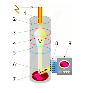 |
| SEM Working flows. |
- Electrons are fired into the machine.
- The main part of the machine (where the object is scanned) is contained within a sealed vacuum chamber because precise electron beams can’t travel effectively through air.
- A positively charged electrode (anode) attracts the electrons and accelerates them into an energetic beam.
- An electromagnetic coil brings the electron beam to a very precise focus, much like a lens.
- Another coil, lower down, steers the electron beam from side to side.
- The beam systematically scans across the object being viewed.
- Electrons from the beam hit the surface of the object and bounce off it.
- A detector registers these scattered electrons and turns them into a picture.
- A hugely magnified image of the object is displayed on a TV screen.
Applications:
- Borders, Forms and Dimensions of Particles.
- Different Phases in a material.
- Composition of different phases by elemental analysis with EDX and WDX spectrometers.
- Micro dimensional impurities by point analysis (EPMA).
Texpedi.com
Check out these related articles:
- Martindale Abrasion Tester Test Results Review
- List of ASTM Testing Standards for Textiles
- Poisson’s Ratio


