Working Principles:
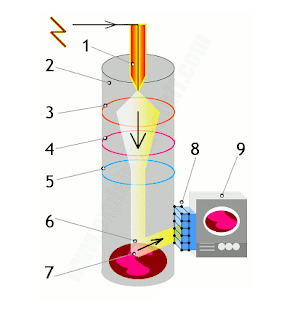
A transmission electron microscope fires a beam of electrons through a specimen to produce a magnified image of an object.
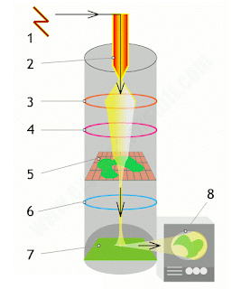 |
| TEM working flows. |
- A high-voltage electricity supply powers the cathode.
- The cathode is a heated filament, a bit like the electron gun in an old-fashioned cathode-ray tube (CRT) TV. It generates a beam of electrons that works in an analogous way to the beam of light in an optical microscope.
- An electromagnetic coil (the first lens) concentrates the electrons into a more powerful beam.
- Another electromagnetic coil (the second lens) focuses the beam onto a certain part of the specimen.
- The specimen sits on a copper grid in the middle of the main microscope tube. The beam passes through the specimen and “picks up” an image of it.
- The projector lens (the third lens) magnifies the image.
- The image becomes visible when the electron beam hits a fluorescent screen at the base of the machine. This is analogous to the phosphor screen at the front of an old-fashioned TV.
- The image can be viewed directly (through a viewing portal), through binoculars at the side, or on a TV monitor attached to an image intensifier (which makes weak images easier to see).
Applications:
- Size of nanoparticles.
- Morphology (structure) of samples.
- Composition and some bonding states.
- Growth of layers, their composition and defects in semiconductors.
- Provides topographical, morphological, compositional and crystalline information.
Texpedi.com
Check out these related articles:





You have shared a nice article here about the Moisture absorber. Your article is very informative and nicely described. I am thankful to you for sharing this article here. moisture absorber manufacturers